Skin health guide · RDG Clinic
Seborrhoeic keratosis assessment in Riga
Seborrhoeic keratosis is usually harmless, but its colour and surface can resemble other lesions. A dermatologist confirms the diagnosis before cosmetic removal.
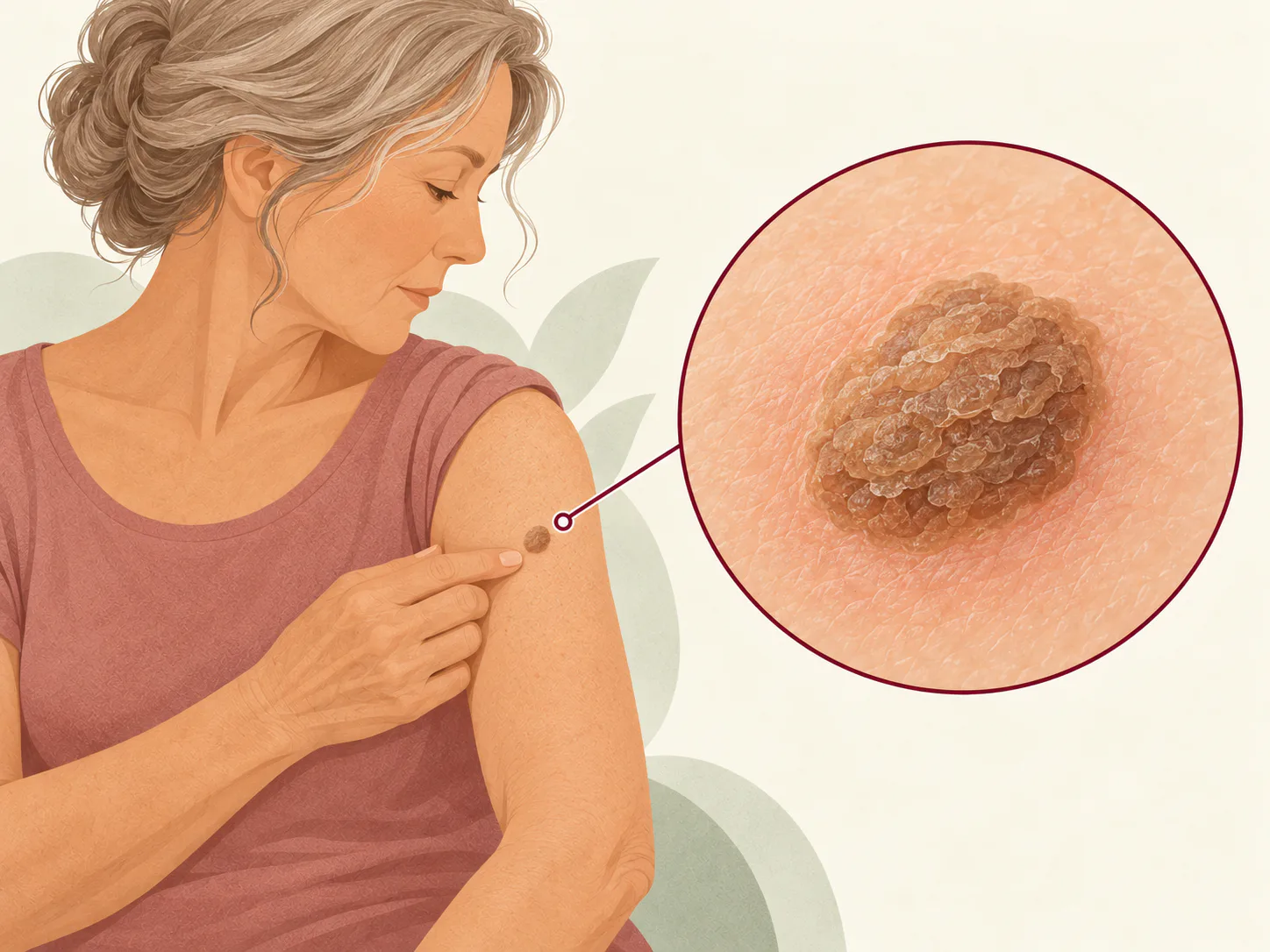
Seborrhoeic keratosis
A benign growth that becomes more common with age
Seborrhoeic keratoses arise in the epidermis and may be light brown, dark, waxy, rough, or appear “stuck on”. They can be single or multiple on the face, trunk, or limbs, but are unusual on palms and soles. They are not contagious and do not turn into melanoma.
However, melanoma, pigmented basal-cell carcinoma, and other lesions can sometimes look similar. A new, rapidly changing, spontaneously bleeding, or distinctly different lesion needs dermoscopic assessment rather than immediate destruction.
Common signs
- A waxy or rough, well-defined surface
- Light brown to almost black colour
- An appearance as if stuck onto the skin
- Itch or irritation from rubbing
When to see a dermatologist
When to see a dermatologist
- The lesion is new or changing rapidly
- It bleeds, ulcerates, hurts, or repeatedly inflames
- You are unsure whether it is keratosis or a mole
- It is repeatedly traumatised or you want removal
Accurate diagnosis
How assessment works
A dermatologist usually recognises typical seborrhoeic keratosis clinically and with dermoscopy. If the structure is atypical or malignancy cannot be confidently excluded, biopsy or complete removal for histology is recommended. A photograph cannot reliably replace in-person examination.
An individual plan
Treatment approach
No treatment is needed when diagnosis is secure and the lesion is asymptomatic. Removal may be considered for symptoms, trauma, or cosmetic preference.
Observation
A typical lesion may be left alone and reviewed if change occurs.
Curettage or coagulation
After anaesthesia, the lesion may be removed mechanically or thermally, with crusting and pigment-change risk.
Biopsy or excision
When diagnosis is uncertain, a method preserving tissue for histology is selected.
Your RDG care path
How the clinic can help
Services are selected after dermatologist assessment and according to the individual findings, not from a name or photograph alone.
Doctor consultation
In person or online: a professional assessment of your health and skin with a clear plan for next steps.
About this service →Skin biopsy and histology
A small tissue sample to clarify diagnosis, using the most suitable biopsy method with or without a stitch.
About this service →Skin lesion removal
Diagnosis first, followed by the most appropriate removal method and histology when required.
About this service →Long-term control
What to expect
After removal, redness, crusting, a lighter or darker mark, and rarely a scar may occur. New seborrhoeic keratoses can arise independently of the previous procedure. Reassessment is needed if the site fails to heal or changes unexpectedly.
This information does not replace medical consultation. Seek urgent medical care for sudden, severe, or rapidly progressive symptoms.
Dermatologists


Frequently asked questions
Can it become cancer?
Seborrhoeic keratosis is benign and does not become melanoma, but another lesion may resemble it.
Is it caused by the sun?
There is no single cause; age and genetics matter, and lesions also occur on covered skin.
Can I scrape it off?
No. This may cause bleeding, infection, scarring, and delay correct diagnosis.
Medical sources
Content updated: 13 October 2026
Treatment starts with an accurate diagnosis
After assessment, the dermatologist will explain the appropriate plan and costs. There is no single fixed treatment price because it depends on the diagnosis and therapy required.